

Bowel, Bladder, Ureter & Diaphragmatic Endometriosis
A patient guide to endometriosis involving organs beyond the usual pelvic surfaces
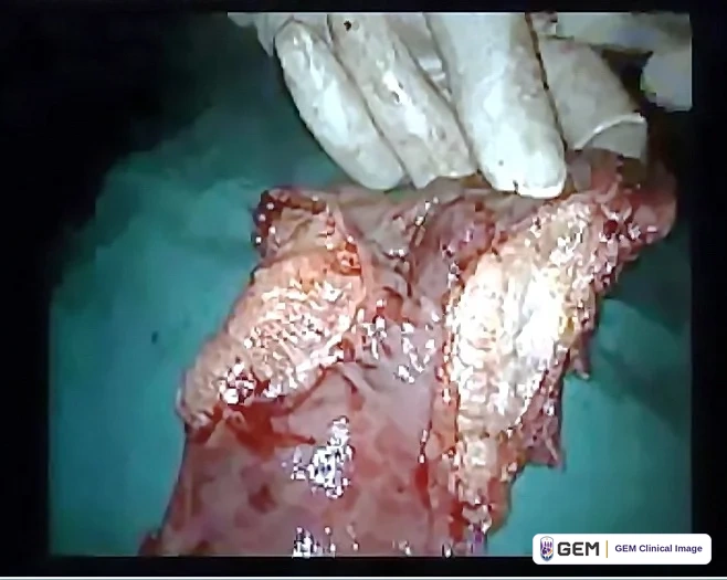
Bowel endometriosis
What it is: Deep endometriosis can infiltrate the bowel wall. Symptoms may include painful bowel movements, cyclical rectal pain, constipation or diarrhea, bloating and pain with sex. Rectal bleeding has several possible causes and needs medical assessment, even if period-related.
Evaluation: Expert ultrasound or MRI may help map disease. Colonoscopy may be needed for other bowel diagnoses but does not reliably exclude bowel endometriosis.
Treatment: Monitoring, hormonal treatment or surgery may be appropriate. Surgery can involve shaving, disc excision or segmental resection, according to symptoms, extent, safety and your preferences. Discuss benefits, bowel-function effects and complications with an experienced team.
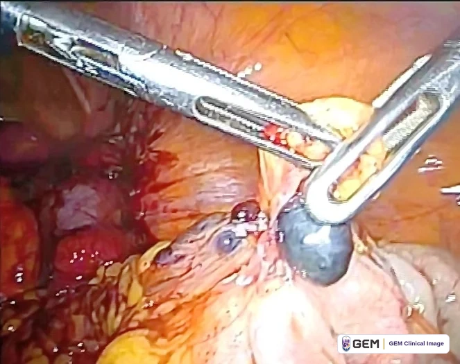
Bladder Endometriosis
What it is: Disease may involve the bladder surface or muscle. Symptoms include cyclical bladder pain, urgency, painful urination and sometimes blood in the urine. Blood in urine needs assessment for other causes too. Expert ultrasound or MRI may help; cystoscopy is useful in selected cases.
Treatment: Monitoring or hormonal treatment may be appropriate when there is no urinary obstruction. Persistent symptoms, obstruction or other clinical concerns may lead to surgery. If bladder-wall resection is needed, an experienced team may perform partial cystectomy and repair, aiming to preserve bladder function and protect the ureteric openings. Decisions depend on disease location, symptoms, fertility goals and patient preferences.
Evidence: A 2022 review reports recurrent urinary symptoms in 7.34% across mainly case reports and series. This is not a lifetime anatomical recurrence rate and does not prove a definitive cure or the superiority of one operation.
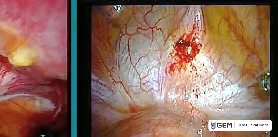
Ureteral Endometriosis
Why it can be silent
Ureteral endometriosis can compress a ureter from outside or, less commonly, involve its wall. It may cause few or no urinary symptoms even when drainage from the kidney is blocked. A normal urinary history does not exclude significant disease.
Silent obstruction can damage kidney function. When ureteral disease is suspected, kidney imaging is important; assess renal function when indicated. Hydronephrosis, or swelling of the kidney from impaired drainage, needs prompt specialist assessment.
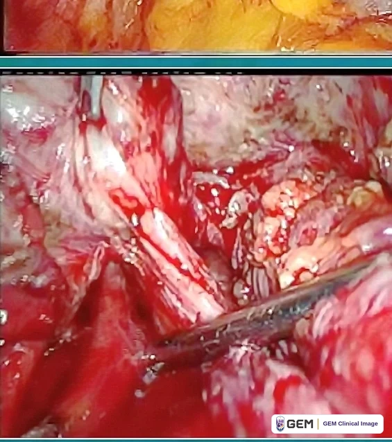
Ureteral Endometriosis – When Suspicion Should Be High
When suspicion should be high
Suspicion is especially important with extensive deep posterior pelvic disease, including rectosigmoid, uterosacral or parametrial involvement. Ureteral disease can coexist with these patterns even without urinary symptoms. Reports from specialist referral populations support careful assessment, but their percentages cannot be applied to every patient.
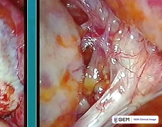
Diaphragmatic & Thoracic Endometriosis
Diaphragmatic disease
Where it occurs: Lesions may occur on the diaphragm, often on the right side, and can coexist with pelvic endometriosis.
Possible symptoms: Cyclical upper abdominal pain, pain beneath the ribs, shoulder-tip pain or pain that worsens around menstruation. Some lesions are asymptomatic and discovered during surgery.
Important point: Diaphragmatic disease may be subtle, small, asymptomatic or hidden, particularly on the posterior right diaphragm behind the liver. Careful inspection is therefore important.
Thoracic disease
What patients should know: Endometriosis involving the chest can present with cyclical chest or shoulder pain, shortness of breath, or catamenial pneumothorax – a collapsed lung occurring in relation to menstruation.
Evaluation: Chest imaging and specialist assessment may be needed; imaging may not identify every lesion.
Team approach: Suspected thoracic disease may require coordinated gynecologic and thoracic surgical expertise; treatment may involve laparoscopy, thoracoscopy, or both.
Preparing for Complex Endometriosis Care
Current major guidelines recommend specialist imaging to assess the extent of suspected deep endometriosis and referral to specialist endometriosis services when disease involves the bowel, bladder or ureter, or occurs outside the pelvis. Complex surgery may require advanced gynecologic laparoscopic expertise together with colorectal, urologic or thoracic surgical expertise.
Questions to ask before complex endometriosis surgery
- Which organs appear to be involved, and how confident are we from imaging?
- Has my imaging been interpreted by someone experienced in deep endometriosis?
- Will a colorectal, urologic or thoracic surgeon be available if needed?
- What type of resection might be necessary and what are the important organ-specific risks?
- Could surgery affect fertility or ovarian reserve?
- Will the surgeon systematically inspect the upper abdomen and diaphragm as well as the pelvis?
- How will disease be mapped, photographed/video-recorded with my consent, described in the operative report and correlated with pathology?
- If all disease cannot safely be resected, what will the follow-up plan be?
Evidence & review
Bowel, Bladder, Ureter & Diaphragmatic Endometriosis
Version 1.1 · Reviewed 13 September 2026
Bowel and bladder treatment
Imaging, symptoms, organ function, fertility plans and patient preferences guide treatment. Bowel surgery can range from shaving to disc or segmental resection in selected patients. For bladder disease without obstruction, monitoring or hormonal treatment may be appropriate. Surgery, including partial cystectomy when indicated, requires attention to bladder function and ureteric openings.
What the bladder review actually reports
Tomasi and colleagues reviewed 28 publications, mainly case reports and series. The abstract reports recurrent urinary symptoms in 7.34%. This is not a lifetime or anatomical lesion-recurrence rate, does not establish that one operation is best, and does not prove a definitive cure. The full paper was not inspected.
Protecting the kidneys
Ureteral disease can be silent and can obstruct drainage from the kidney. When ureteral involvement is suspected, assess the upper urinary tract with kidney imaging and evaluate renal function when indicated. Hydronephrosis warrants prompt specialist assessment. Published referral cohorts and case reports establish the possibility of silent harm; they do not supply a single risk percentage for every patient.
Interpreting the removed percentages
The original approximate 70% and 90% figures reflect selected referral-series findings. Alborzi reports 353 enrolled and 326 pathology-confirmed patients, with 70.53% rectosigmoid and 92.9% uterosacral involvement. These are not general-population estimates. The 10-14% statement is background in a case report, not a universal probability. The corrected patient text uses the clinically relevant qualitative warning.
Diaphragm, chest and safe surgical planning
Assess suspected chest involvement with appropriate specialists. Sudden chest pain or serious breathing difficulty requires emergency assessment regardless of menstrual timing. A safe, systematic survey documents accessible visible surfaces; it does not guarantee that all lesions are visible or require automatic removal of incidental asymptomatic disease. Treatment may be staged according to consent, safety and available expertise.
Medical information
Seek urgent or emergency assessment for severe or rapidly worsening symptoms.
Version 1.1 · Reviewed 13 September 2026. Proposed next review: September 2027, or sooner if relevant guidance changes.
Full references
Numbers correspond to the sources cited in this revised guide. Access notes distinguish relevant full-text inspection from abstract-only checks. No source is presented as an endorsement of GEM.
- [1] ESHRE. Endometriosis guideline. 2022.
Verification: Relevant full-guideline sections inspected online.
Freely available — https://www.eshre.eu/-/media/sitecore-files/Guidelines/Endometriosis/ESHRE-GUIDELINE-ENDOMETRIOSIS-2022_1.pdf - [2] Working group of ESGE, ESHRE, and WES, et al. Recommendations for the surgical treatment of endometriosis. Part 2: deep endometriosis. Hum Reprod Open. 2020;2020(1):hoaa002. doi:10.1093/hropen/hoaa002. PMID 32064361.
Verification: Bibliographic record, abstract and relevant full-text sections inspected via Europe PMC.
Freely available — https://europepmc.org/articles/PMC7013143 - [3] Leonardi M, Espada M, Kho RM, et al. Endometriosis and the Urinary Tract: From Diagnosis to Surgical Treatment. Diagnostics (Basel). 2020;10(10):E771. doi:10.3390/diagnostics10100771. PMID 33007875.
Verification: Bibliographic record, abstract and relevant full-text sections inspected via Europe PMC.
Freely available — https://europepmc.org/articles/PMC7650710 - [4] Tomasi MC, Ribeiro PAA, Farah D, et al. Symptoms and Surgical Technique of Bladder Endometriosis: A Systematic Review. J Minim Invasive Gynecol. 2022;29(12):1294-1302. doi:10.1016/j.jmig.2022.10.003. PMID 36252916.
Verification: Bibliographic record and complete abstract inspected via PubMed/Europe PMC; full paper not inspected.
Freely available — https://pubmed.ncbi.nlm.nih.gov/36252916/ - [5] Barra F, Scala C, Biscaldi E, et al. Ureteral endometriosis: a systematic review of epidemiology, pathogenesis, diagnosis, treatment, risk of malignant transformation and fertility. Hum Reprod Update. 2018;24(6):710-730. doi:10.1093/humupd/dmy027. PMID 30165449.
Verification: Bibliographic record and complete abstract inspected via PubMed/Europe PMC; full paper not inspected.
Freely available — https://pubmed.ncbi.nlm.nih.gov/30165449/ - [6] Muñoz JL, Jiménez JS, Tejerizo A, et al. Rectosigmoid deep infiltrating endometriosis and ureteral involvement with loss of renal function. Eur J Obstet Gynecol Reprod Biol. 2012;162(2):121-124. doi:10.1016/j.ejogrb.2012.02.030. PMID 22534016.
Verification: Bibliographic record and complete abstract inspected via PubMed/Europe PMC; full paper not inspected.
Freely available — https://pubmed.ncbi.nlm.nih.gov/22534016/ - [7] Cavaco-Gomes J, Martinho M, Gilabert-Aguilar J, et al. Laparoscopic management of ureteral endometriosis: A systematic review. Eur J Obstet Gynecol Reprod Biol. 2017;210:94-101. doi:10.1016/j.ejogrb.2016.12.011. PMID 27984749.
Verification: Bibliographic record and complete abstract inspected via PubMed/Europe PMC; full paper not inspected.
Freely available — https://pubmed.ncbi.nlm.nih.gov/27984749/ - [8] Alborzi S, Askary E, Poordast T, et al. Approach to ureteral endometriosis: A single-center experience and meta-analysis of the literature. J Obstet Gynaecol Res. 2023;49(1):75-89. doi:10.1111/jog.15449. PMID 36268633.
Verification: Bibliographic record and complete abstract inspected via PubMed/Europe PMC; full paper not inspected.
Freely available — https://pubmed.ncbi.nlm.nih.gov/36268633/ - [9] Luzny R, Herman H, Malchar O, et al. Hydronephrosis as a symptom of clinically silent ureteral endometriosis. Ceska Gynekol. 2022;87(3):188-192. doi:10.48095/cccg2022188. PMID 35896397.
Verification: Bibliographic record and complete abstract inspected via PubMed/Europe PMC; full paper not inspected.
Freely available — https://pubmed.ncbi.nlm.nih.gov/35896397/ - [10] Naem A, Roman H, Martin DC, Krentel H. A bird-eye view of diaphragmatic endometriosis: current practices and future perspectives. Front Med. 2024;11:1505399. doi:10.3389/fmed.2024.1505399.
Verification: Publisher article and indexed abstract inspected.
Freely available — https://www.frontiersin.org/journals/medicine/articles/10.3389/fmed.2024.1505399/full - [11] NHS. Laparoscopy (keyhole surgery). Reviewed 20 December 2023.
Verification: Full patient page inspected.
Freely available — https://www.nhs.uk/tests-and-treatments/laparoscopy/
This material is for educational purposes only and does not constitute medical advice or create a clinician-patient relationship with GEM.
© 2026 Global Endometriosis Movement. All rights reserved.