

What Endometriosis Can Actually Look Like
Illustrative surgical appearances and locations; photographs alone do not confirm a diagnosis.
1. Normal pelvis
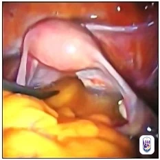
A reference view of normal pelvic anatomy, including the ovary and fallopian tube.
2. Superficial endometriosis: red appearance
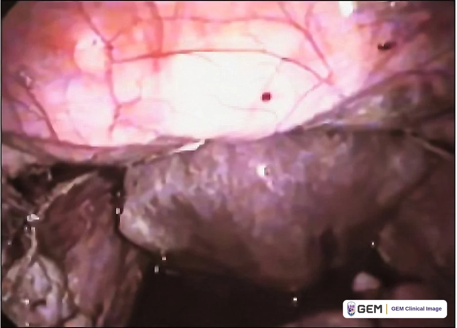
Red-appearing lesion. Color alone cannot establish a lesion's age, biological activity or diagnosis.
3. Superficial endometriosis: dark appearance
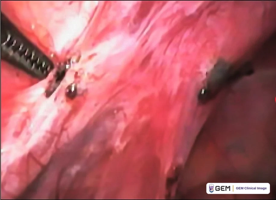
Superficial implants may also appear dark brown or black rather than red.
4. Ovarian endometrioma
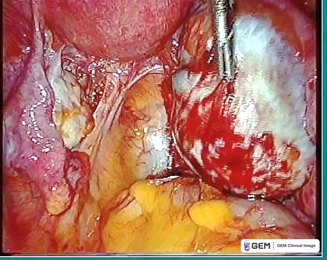
Endometriosis involving the ovary. These cysts contain old blood and are often called 'chocolate cysts'.
5. Deep endometriosis
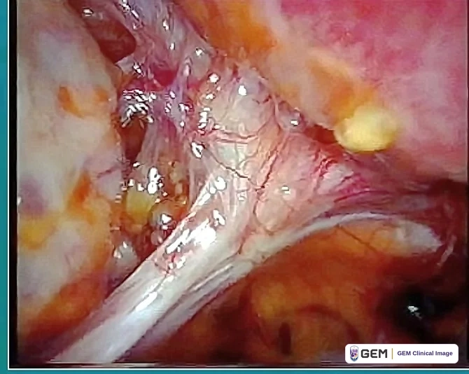
Deep disease extends beneath the peritoneal surface and may cause fibrosis, scarring and distorted anatomy.
6. Bladder endometriosis
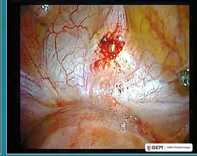
Deep endometriosis can involve the bladder wall – an example of disease beyond the reproductive organs.
Deep Endometriosis Can Involve Other Organs
Authentic surgical photographs demonstrating ureteral and bowel involvement
7. Ureter + large-bowel involvement
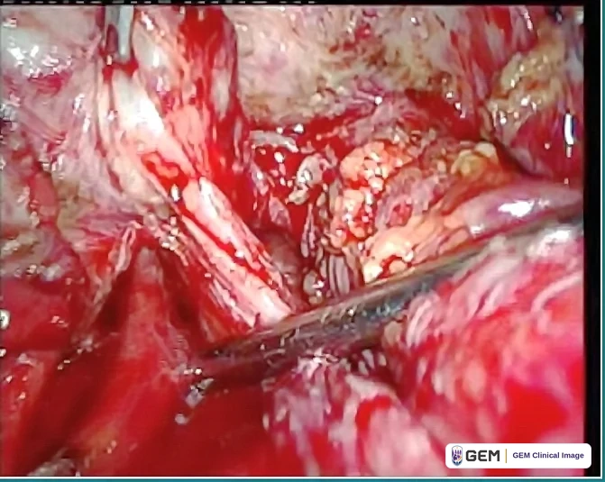
Deep endometriosis can involve structures close together in the pelvis, including the ureter and large bowel.
8. Cut section of large bowel
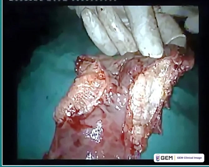
A cut bowel specimen, labeled as deep endometriosis in the supplied file. Extent and diagnosis require correlation with the operative and pathology records.
9. Large-bowel endometriosis
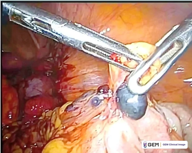
Another operative view of bowel involvement, illustrating the anatomical complexity of deep endometriosis.
These photographs show different phenotypes and locations – not an inevitable step-by-step progression.
How to Read These Photographs
A short patient explanation and scientific context
Three major forms are commonly recognized
Endometriosis can present as superficial peritoneal disease, ovarian endometrioma ('chocolate cyst'), and deep endometriosis. More than one form can occur in the same patient.
Deep disease can involve organs
Deep endometriosis extends beneath the peritoneal surface and may affect the uterosacral ligaments, pelvic sidewall, vagina, bowel, bladder or ureter.
Appearance is not the same as symptoms
A visually small amount of disease can be associated with substantial pain, while extensive disease may sometimes produce fewer symptoms. Conventional stage does not reliably predict pain or surgical complexity.
How to interpret the captions
The sequence shows different appearances and anatomical locations, not an inevitable progression or pain-severity scale. The supplied clinical labels must be checked against the original operative/pathology records before publication.
The photographs are educational examples, not a diagnostic test. Color alone does not establish lesion age or activity.
Evidence & references
What Endometriosis Actually Looks Like
Version 1.1 · Reviewed 13 September 2026
Appearances and limitations
Superficial, ovarian and deep disease can coexist. Color, visible extent and conventional stage do not reliably determine pain, lesion age or activity. Photographs cannot replace clinical assessment or pathology.
Case labels remain provisional
The nine images and their layout are retained. Their labels describe the supplied intended examples, not independently verified diagnoses. Match each image to its source operation/pathology before public use. The series is not a step-by-step progression scale.
Full references
- [S01] ESHRE. Endometriosis guideline. 2022. Freely available
- [S11] Working group of ESGE, ESHRE and WES, Saridogan E, et al. Recommendations for the Surgical Treatment of Endometriosis. Part 1: Ovarian Endometrioma. Hum Reprod Open. 2017;2017(4):hox016. doi:10.1093/hropen/hox016. PMID 31486802. Freely available
- [S12] Working group of ESGE, ESHRE, and WES, et al. Recommendations for the surgical treatment of endometriosis. Part 2: deep endometriosis. Hum Reprod Open. 2020;2020(1):hoaa002. doi:10.1093/hropen/hoaa002. PMID 32064361. Freely available
- [R34693033] International Working Group of AAGL, ESGE, ESHRE and WES et al. An international terminology for endometriosis, 2021. Hum Reprod Open. 2021;2021(4):hoab029. doi:10.1093/hropen/hoab029. PMID 34693033. Freely available
No listed institution endorses GEM.
Medical information
Do not delay care; seek emergency help for a suspected emergency.
This material is for educational purposes only and does not constitute medical advice or create a clinician-patient relationship with GEM.
© 2026 Global Endometriosis Movement. All rights reserved.