

Endometriosis & Adenomyosis
They can cause similar symptoms, but they are not the same condition.
How are they found?
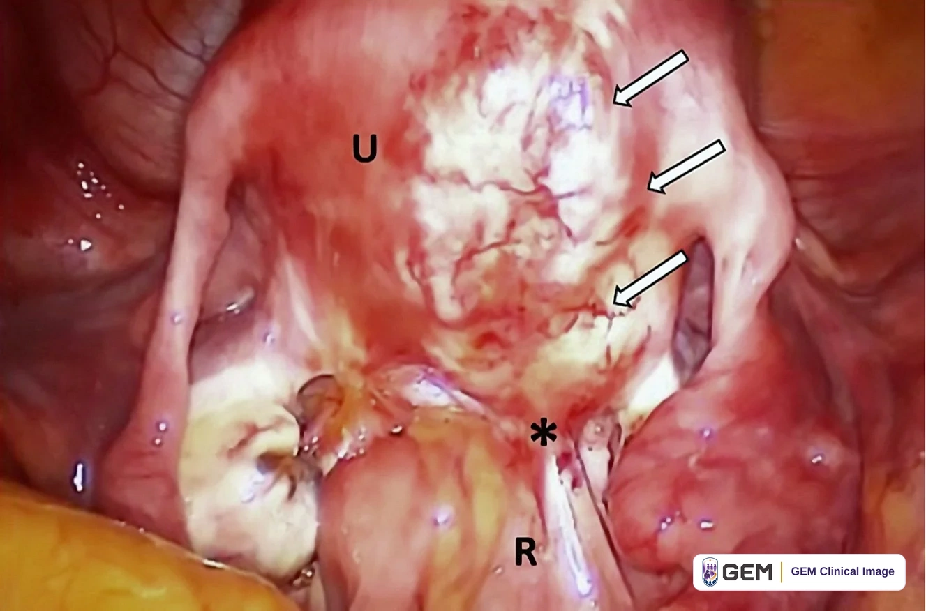
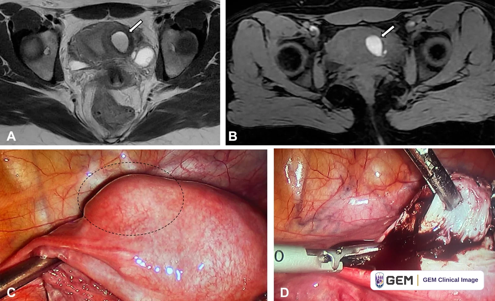
Symptoms, examination, ultrasound and sometimes MRI help assessment. External uterine appearance alone cannot diagnose adenomyosis. Normal imaging does not exclude all endometriosis.
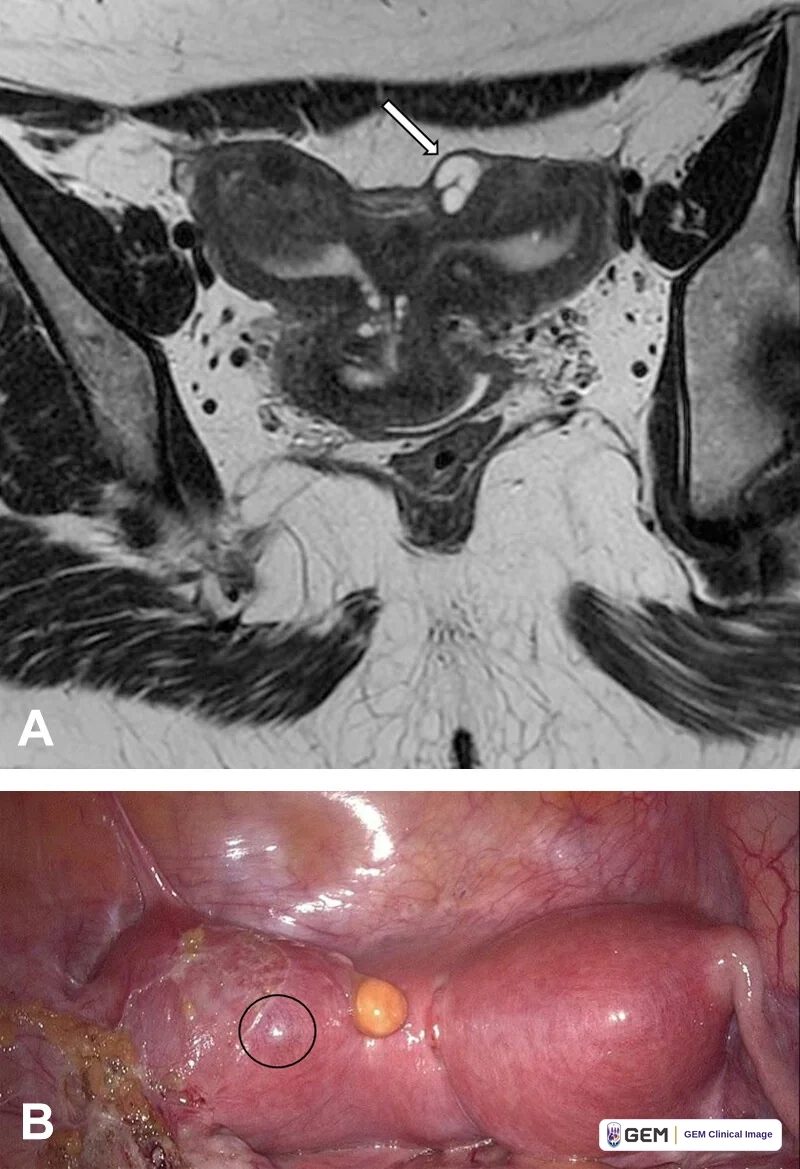
Why Does the Difference Matter?
Because the condition causing your symptoms can change the treatment you need.
How is adenomyosis treated?
Treatment depends on symptoms, age and reproductive goals. Medicines can help pain and bleeding; fertility treatment may be appropriate. Selected uterus-preserving surgery requires expert counselling about benefits, complications and risks to a future pregnancy. No single treatment suits everyone.
What about hysterectomy?
Hysterectomy treats uterine adenomyosis; you cannot carry a pregnancy afterward. Endometriosis elsewhere or other causes of pain may remain.
Evidence & review
Endometriosis & Adenomyosis
Version 1.3 · Reviewed 13 September 2026
Definitions and diagnosis
Endometriosis is endometrium-like tissue outside the uterine endometrium and myometrium. Adenomyosis involves endometrial glands and stroma within the myometrium, the muscular uterine wall. They can coexist. Ultrasound and MRI can support diagnosis; external laparoscopic appearance alone does not establish adenomyosis.
Coexistence is an association
Marcellin and colleagues studied 255 symptomatic patients undergoing surgery for deep endometriosis. The association with focal outer-myometrial adenomyosis is a finding in a selected surgical population, not proof that endometriosis causes adenomyosis or that all patients have both.
Fertility evidence has limits
A 2024 IVF/ICSI meta-analysis associates adenomyosis with lower pregnancy and live-birth rates and higher miscarriage rates. The evidence is largely observational and concerns assisted reproduction; it is not an individual prediction or direct estimate for all natural conceptions. Newer reviews describe heterogeneity and uncertain treatment effects.
Treatment and hysterectomy
Treatment depends on symptoms and reproductive goals. Medicines can help pain and bleeding. Selected uterus-preserving surgery needs specialist counselling, including effects on future pregnancy. Hysterectomy definitively treats adenomyosis in the removed uterus and prevents carrying a pregnancy. It does not remove endometriosis elsewhere or guarantee that every cause of pelvic pain will resolve.
Medical information
Seek urgent or emergency assessment for severe or rapidly worsening symptoms.
Version 1.3 · Reviewed 13 September 2026. Proposed next review: September 2027, or sooner if relevant guidance changes.
Full references
Numbers correspond to the sources cited in this revised guide. Access notes distinguish relevant full-text inspection from abstract-only checks. No source is presented as an endorsement of GEM.
- [1] ESHRE. Endometriosis guideline. 2022.
Verification: Relevant full-guideline sections inspected online.
Freely available — https://www.eshre.eu/-/media/sitecore-files/Guidelines/Endometriosis/ESHRE-GUIDELINE-ENDOMETRIOSIS-2022_1.pdf - [2] NHS. Adenomyosis. Reviewed 17 July 2023.
Verification: Relevant patient page inspected.
Freely available — https://www.nhs.uk/conditions/adenomyosis/ - [3] Marcellin L, Santulli P, Bourdon M, et al. Focal adenomyosis of the outer myometrium and deep infiltrating endometriosis severity. Fertil Steril. 2020;114(4):818-827. doi:10.1016/j.fertnstert.2020.05.003. PMID 32741618.
Verification: Bibliographic record and complete abstract inspected via PubMed/Europe PMC; full paper not inspected.
Freely available — https://pubmed.ncbi.nlm.nih.gov/32741618/ - [4] Ge L, Li Y, Zhou J, et al. Effect of different treatment protocols on in vitro fertilisation/intracytoplasmic sperm injection (IVF/ICSI) outcomes in adenomyosis women: a systematic review and meta-analysis. BMJ Open. 2024;14(7):e077025. doi:10.1136/bmjopen-2023-077025. PMID 39025820.
Verification: Bibliographic record, abstract and relevant full-text sections inspected via Europe PMC.
Freely available — https://europepmc.org/articles/PMC11261672 - [5] Kolovos G, Kalampokas T, Dedes I, et al. The impact of adenomyosis localization in myometrium on fertility and pregnancy outcomes: a narrative systematic review of the literature. Front Reprod Health. 2026;8:1748474. doi:10.3389/frph.2026.1748474. PMID 41918520.
Verification: Bibliographic record and complete abstract inspected via PubMed/Europe PMC; full paper not inspected.
Freely available — https://pubmed.ncbi.nlm.nih.gov/41918520/ - [6] Manem N, Hori K, Aghajanova L. Adenomyosis and fertility: from pathophysiology to optimizing reproductive outcomes. Curr Opin Obstet Gynecol. 2026;38(4):208-216. doi:10.1097/gco.0000000000001117. PMID 42244463.
Verification: Bibliographic record and complete abstract inspected via PubMed/Europe PMC; full paper not inspected.
Freely available — https://pubmed.ncbi.nlm.nih.gov/42244463/ - [7] Capezzuoli T, Toscano F, Ceccaroni M, et al. Conservative surgical treatment for adenomyosis: New options for looking beyond uterus removal. Best Pract Res Clin Obstet Gynaecol. 2024;95:102507. doi:10.1016/j.bpobgyn.2024.102507. PMID 38906739.
Verification: Bibliographic record and complete abstract inspected via PubMed/Europe PMC; full paper not inspected.
Freely available — https://pubmed.ncbi.nlm.nih.gov/38906739/
This material is for educational purposes only and does not constitute medical advice or create a clinician-patient relationship with GEM.
© 2026 Global Endometriosis Movement. All rights reserved.