

Getting the Diagnosis
From clinical suspicion to imaging, definitive surgical assessment, treatment and documentation
Diagnosis begins with the patient's history and a careful clinical assessment. Imaging is extremely valuable, but it has limitations. GEM believes patients should understand both the strengths of modern imaging and the unique diagnostic-and-therapeutic role of expertly performed laparoscopy when surgery is appropriate.
1. Why diagnosis is often delayed

Severe menstrual pain may be dismissed or attributed to other causes. Start with a careful history. An examination may help, but its purpose, consent and comfort should be discussed; an internal examination is not mandatory for every patient.
2. Preparing for your doctor's visit

Write down your symptoms before the appointment. Note when they occur, whether they change around your period, their severity, and how they affect school, work, sleep, exercise, sex, bowel or bladder function, and fertility. Bring previous imaging, operative reports, medications and relevant family history.
3. Medical history and examination

Your history is a major part of the diagnosis. Physical and pelvic examination may identify tenderness, an ovarian mass, reduced organ mobility, nodularity or findings suggesting deep disease. A normal examination, however, does not rule out endometriosis.
4. Ultrasound
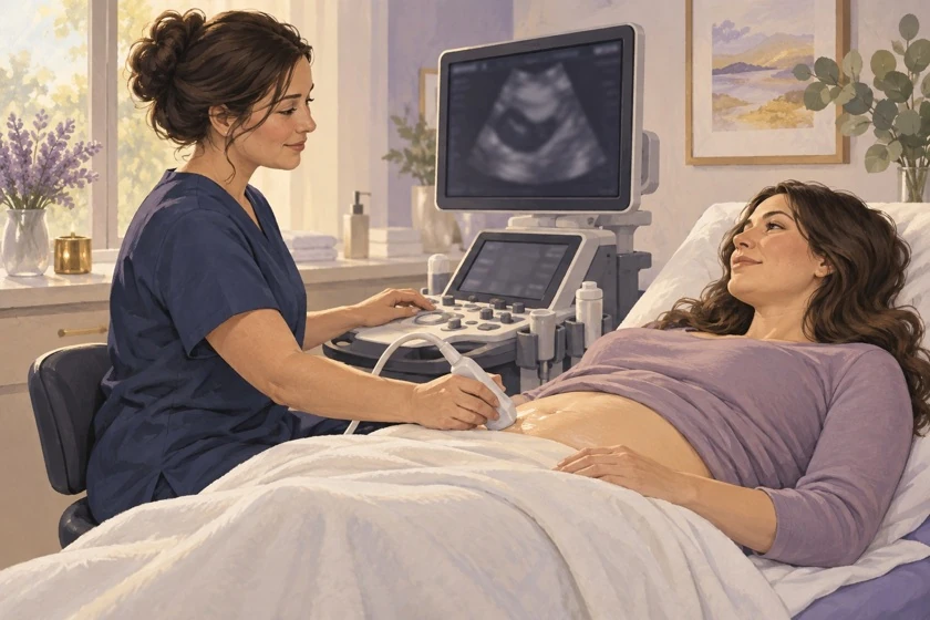
Ultrasound is a first-line imaging option. Transvaginal ultrasound can identify endometriomas and some deep disease when acceptable and suitable. If it is declined or unsuitable, discuss transabdominal ultrasound or other appropriate imaging. Expertise matters.
5. MRI
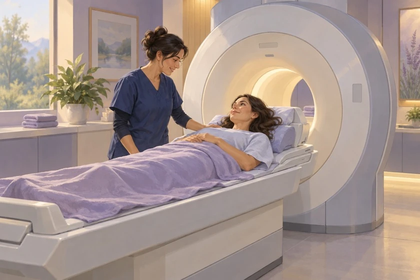
MRI is not required routinely for every patient. It may be useful when deep or anatomically complex disease is suspected, ultrasound is uncertain, or additional preoperative mapping is needed. MRI complements – rather than replaces – clinical assessment and appropriate ultrasound.
6. What a normal ultrasound does – and does not – mean

A normal ultrasound does not mean that you do not have endometriosis. Small superficial peritoneal lesions may not be visible. When the clinical picture remains strongly compatible with endometriosis, a normal scan should not automatically end the evaluation.
7. Is there a blood test?

CA-125 should not be used alone to diagnose or exclude endometriosis. New biomarker tests are being evaluated; their evidence and availability vary. A test result should be interpreted within the current local diagnostic pathway.
8. When is laparoscopy appropriate?

When clinical suspicion and preoperative evaluation provide sufficient reason for surgery, laparoscopy should be carefully planned. GEM's objective is not a diagnostic-only operation followed by another surgery because the first team was not prepared. Unexpected findings, safety concerns, limits of consent or unanticipated organ involvement may occasionally justify staging.
9. Why complete video documentation matters

With the patient's informed consent, GEM believes endometriosis surgery should be comprehensively documented, including preservation of the complete unedited operative video whenever feasible and consistent with privacy, institutional and legal requirements.
10. Questions after diagnosis

Ask: What supports my diagnosis? What might imaging miss? What medical and surgical options fit my goals? If surgery is advised, what areas can be safely assessed, what treatment is consented, and what findings, pathology, photographs or video will be available?
Diagnosis and follow-up
Normal imaging does not exclude all endometriosis, especially superficial disease. Symptom improvement with hormones does not prove the diagnosis or prove that lesions have disappeared. Reassessment should follow symptoms, treatment response and any concern about organ involvement.
GEM's preference for comprehensive mapping and complete unedited operative video is an advocacy position. Recording depends on informed consent, feasibility and applicable privacy requirements. It is not a universal legal duty.
Evidence & references
Getting the Diagnosis
Version 1.2 · September 2026
Diagnostic pathway
Clinical history, consensual examination and suitable imaging guide care. Normal imaging does not exclude superficial disease. Empirical treatment and laparoscopy are individualized options, not a compulsory sequence for everyone.
Tests and surgical documentation
CA-125 cannot diagnose/exclude endometriosis alone. Emerging tests require current local guidance; no new commercial test is endorsed here. GEM's preference for therapeutic planning and complete video is labeled advocacy, not a universal guideline or legal requirement.
Full references
- [S01] ESHRE. Endometriosis guideline. 2022. Freely available
- [S13] WHO. Endometriosis. Fact sheet, 15 October 2025. Freely available
- [R34693033] International Working Group of AAGL, ESGE, ESHRE and WES et al. An international terminology for endometriosis, 2021. Hum Reprod Open. 2021;2021(4):hoab029. doi:10.1093/hropen/hoab029. PMID 34693033. Freely available
No listed institution endorses GEM.
Medical information
Do not delay care; seek emergency help for a suspected emergency.
This material is for educational purposes only and does not constitute medical advice or create a clinician-patient relationship with GEM.
© 2026 Global Endometriosis Movement. All rights reserved.